Product code
A10206
Name of the product
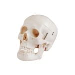
Skull and Brain Model 8 Pieces


Product description
This model is designed for visual aid in teaching physiology and health information lessons. It helps students understand morphology, skull and brain formation.
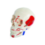
It consists of 22 bones with jagged stitch lines marked and the brain consists of 8 parts.
Skull: The calvarium is cut transversely to show the internal structures of the skull. The lower jaw is mobile, 3 lower incisors, canines and molars can be removed.
External Features of the Brain:
- Cerebral hemisphere; On the dossolateral surface, the central sulcus and lateral and parietooccipital fissures, frontal, parietal, temporal and posterior head lobes are shown. On the median and basal surface, the cracks of the main callos (cutting surface), parietooccipital and limestone, the nasal bulb and its paths are shown.
- Brainstem; On the dorsal aspect, the thalamus, pulvinar quadruplet hillock and rhomboid fossa are shown. On the abdominal side, optic schizma, mammillary organs, crura pedicle and bridges are depicted.
- Cerebellum; Other features of the cerebellar hemispheres and lobes are marked.
Artery Supply of the Brain:
- Sources: 2 vertebral 2 internal carotid arteries.
- The right and left vertebral veins pass forward from the ventral surface of the bulbus, joining at the lower border of the bridges to form unpaired basilar arteries. At the upper border of the bridges, after giving a pair of hindbrain arteries, it divides the hindbrain arteries. The hindbrain is connected to the inner right and left carotid arteries by previous branches of communication. Internal carotid leaves the middle and forebrain arteries.
- The cerebellum and artery supply of the brain.
- The cerebellum is supplied from 3 pairs of cerebral arteries.
- Post inferior brain arteries arise from vertebral arteries.
- The anterior inferior cerebral arteries originate from the basilar artery at the lower border of the bridges.
- The posterior cerebral arteries originate from the basilar artery at the more posterior border of the bridges.
- The cerebellum is generally supplied from 3 pairs of cerebral arteries.
- Posterior brain arteries, end branches of the basilar arteries.
- From the lateral fissure roots the midbrain arteries supply the main part of the dorsal lateral surface of the brain.
- The forebrain arteries are the end branches of the medial surface of the internal carotid arteries in the brain hemispheres.
- The cerebellum is supplied from 3 pairs of cerebral arteries.
(The vesele passing between the dorsal surface of the thalamus and the groove in the caudate nucleus is called the vein, vena terminalis).
MATERIAL
The model is made of PVC plastic.
DIMENSIONS
Real size.
WEIGHT
- Gross: 1,3 kg
- Net: 1.2 kg